
![]()

|
GENTAUR EUROPE BELGIUM1 tel +32 2 732 5688 fax +32 2 732 4414 [email protected] Av. de l' Armée 68 B-1040 Brussels France tel 01 43 25 01 50 fax 01 43 25 01 60 9, rue Lagrange 75005 Paris Italy tel 02 36 00 65 93 fax +32 16 50 90 45 20135 Milano Germany tel +32 16 58 90 45 fax +32 16 50 90 45 Forckenbeckstraße 6 D-52074 Aachen Japan tel +81 78 386 0860 fax +81 78 306 0296 Minaatojimaminami-manchi Chuo-ku, Kobe 065-0047 |
| Carboxyfluorescein FLICA Assays for Detection of
Caspase Activity
The central component of apoptosis is a cascade of proteolytic enzymes called caspases. These enzymes participate in a series of reactions that are triggered in response to pro-apoptotic signals and result in the cleavage of protein substrates, causing the disassembly of the cell1. FLICA Apoptosis Detection Kits use a novel approach to detect active caspases. The methodology is based on a Fluorochrome Inhibitor of Caspases (FLICA). Once inside the cell, the FLICA inhibitor binds covalently to the active caspase2. These inhibitors are cell permeable and non-cytotoxic. For detection using green fluorescence, a carboxyfluorescein-labeled fluoromethyl ketone peptide inhibitor of caspases is used.
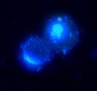
In Figure 1, photo A, only one cell appears green it is apoptotic and stained positive for poly caspase activity with the FAM-VAD-FMK reagent. The other cell, which is not visible, did not bind to the reagent and therefore is not apoptotic. The same cells, photographed at right under a different wavelength for Hoechst stain, appear blue. The cell in the top right of photo B (which appears green in photo A) has a very brightly stained nucleus its DNA is condensing in the cell, a sign it is dying. The cell in the bottom left of photo B (which is not visible in photo A) does not have a brightly stained nucleus, therefore it is neither apoptotic nor necrotic. References: |
||||||
![]()
![]()